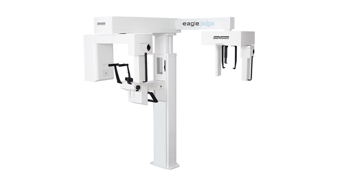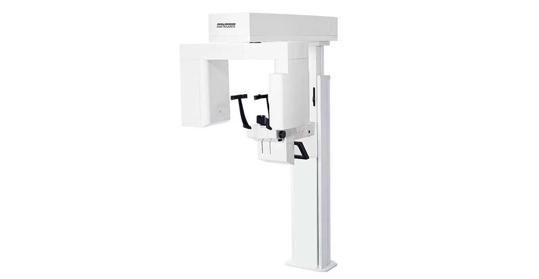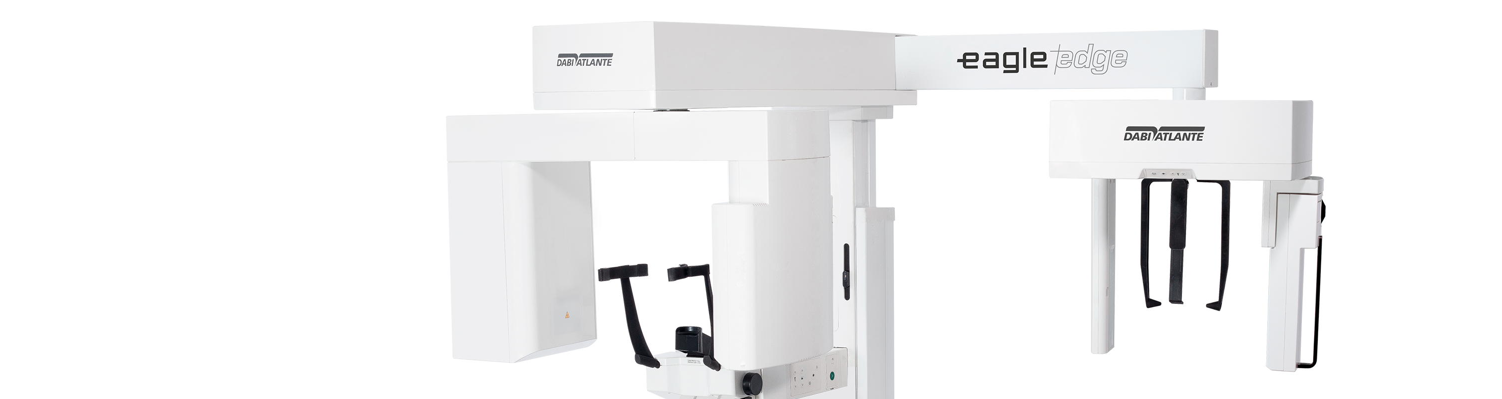
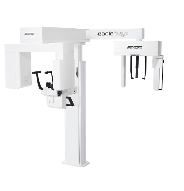
Eagle Edge line
V-BEAM – Variable Cone-Beam
Variable Cone Beam, exclusive technology developed by Eagle, guarantees high definition in images with FOV of 5×5Ø, 6×9Ø and 9×9Ø as well as allowing the capture of larger images. Eagle Edge is the complete solution for three-dimensional diagnostics, especially in endodontic, implant dentistry and orthodontic applications.
6 Tomographic volumes
5×5 – Endo | 6×9 – Upper or Lower Jaw | 9×9 – Full Jaw | 9×16 – Extended Jaw | 15×16* – Skull | 21×16*: Skull
*Volume built from fusion (Auto Stitching)
85µm ultra high resolution
Eagle Edge has different resolutions with Isotropic Voxel between 85 and 400 μm, with automatic adjustment in relation to volume size and resolution.
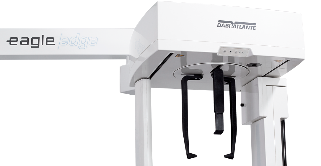
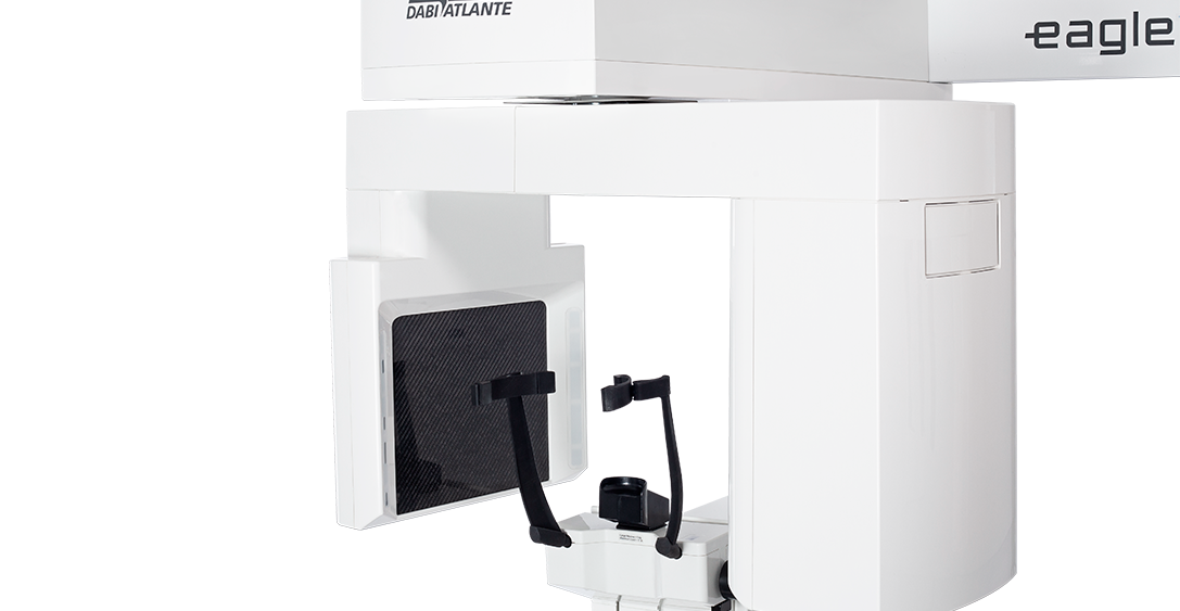
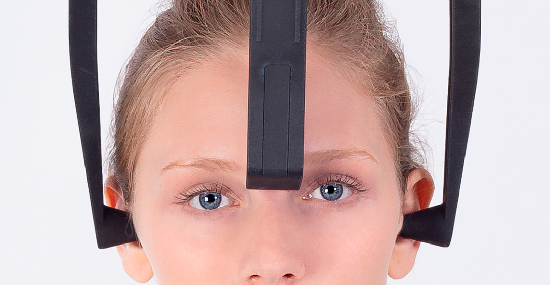
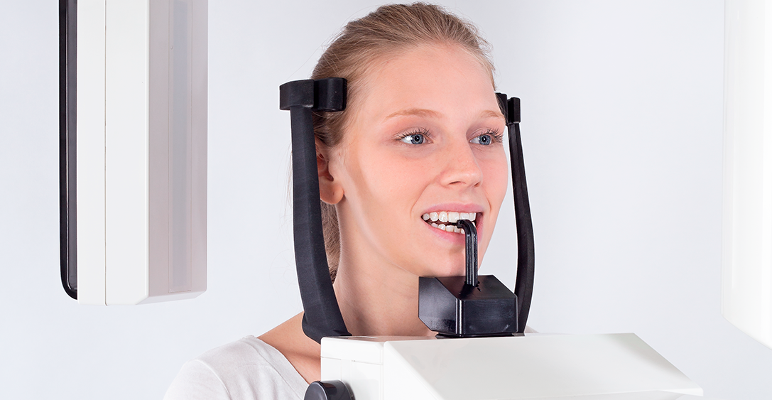
Stability and usability
Eagle Edge has new head positioners with 4 support points for better patient stability during exams. Simple to use and easy to position, the set of head positioners was designed to facilitate the clinical routine, making the execution in sequence faster.
3 axis movement
The latest generation movement system that includes three axes (two orthogonal directions and one rotation) which allows greater flexibility in the elaboration of radiographic profiles, optimization of the thickness of the cutting plane and constant vertical magnification.
Eagle Smart Contrast
Innovative algorithm that works in all regions of the image, treating and improving the contrast of each area individually. The result is a homogeneous and noise-free image, allowing the visualization of details and, consequently, better diagnosis.
DICOM Server
The Dicom Send tool of the Eagle software allows the instantaneous sending of the images generated by the equipment to image storage and sharing systems in physically different locations.
Reduction of metallic artifacts
Eagle Edge features options with selectable processing levels to correct deformations of gutta-percha, implants and/or wide prostheses and metal restorations, in addition to automatic metal reduction. This feature also allows image reprocessing for a better diagnosis without the need to generate new exposure to the patient.
Eagle Eye
During the performance of a panoramic radiograph, hundreds of images are generated and assembled into a final image. Eagle Eye software features an innovative function (algorithm) that scans all processed images, seeking the best definition of focus in order to deliver a final image with greater detail and definition, especially in the region of the incisors and canines, ATM and root canals.