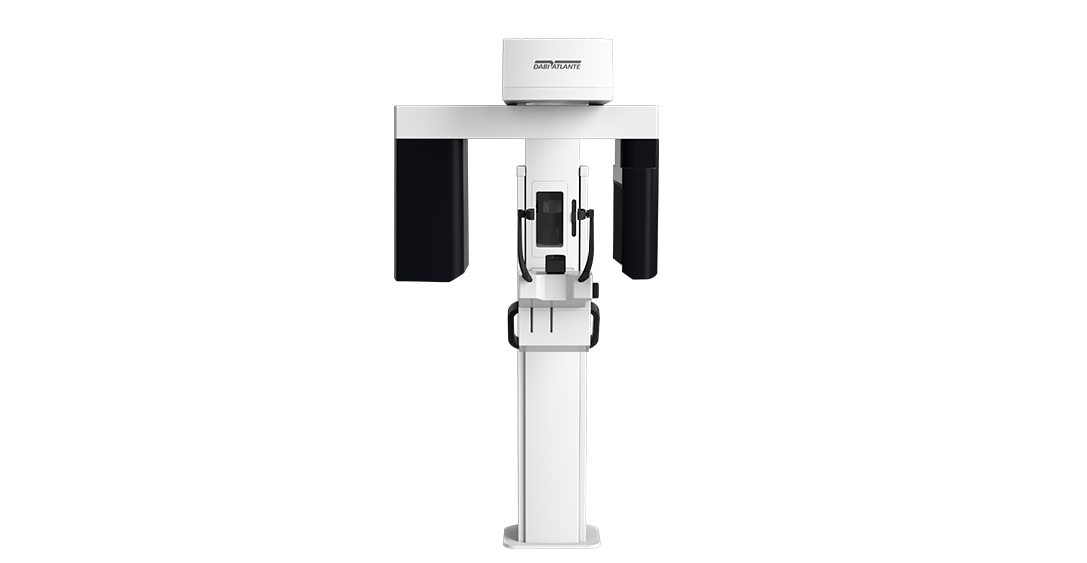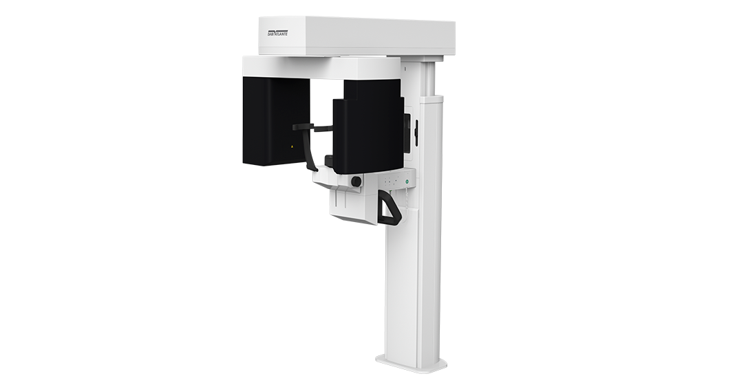
Eagle Edge 0.2 FS
High definition images:
- Unique with 0.2 focal point, which generates stunning images
- Horizontal x-ray beams to reduce metallic artifacts
- 360º scanning
- Sensor with high quantum efficiency
PATIENT MOTION CORRECTION
During the exams, the micro-movement of the patient is common, so that the final result of the exam can be harmed.
The Eagle Edge algorithm automatically corrects the image, ensuring the best Quality of the exam, avoiding repetitions and offering greater acuity for performing diagnoses.
V-BEAM – Variable Cone-Beam
Variable Cone Beam, exclusive technology developed by Eagle, guarantees high definition in images with FOV of 5×5Ø, 6×9Ø and 9×9Ø as well as allowing the capture of larger images. Eagle Edge is the complete solution for three-dimensional diagnostics, especially in endodontic, implant dentistry and orthodontic applications.
3 Tomographic volumes
5×5 – Endo | 6×9 – Upper or Lower Jaw | 9×9 – Complete Jaw
75µm ultra high resolution
Eagle Edge 0.2 FS has different resolutions with Isotropic Voxel between 75 and 200 μm, with automatic adjustment in relation to volume size and resolution.
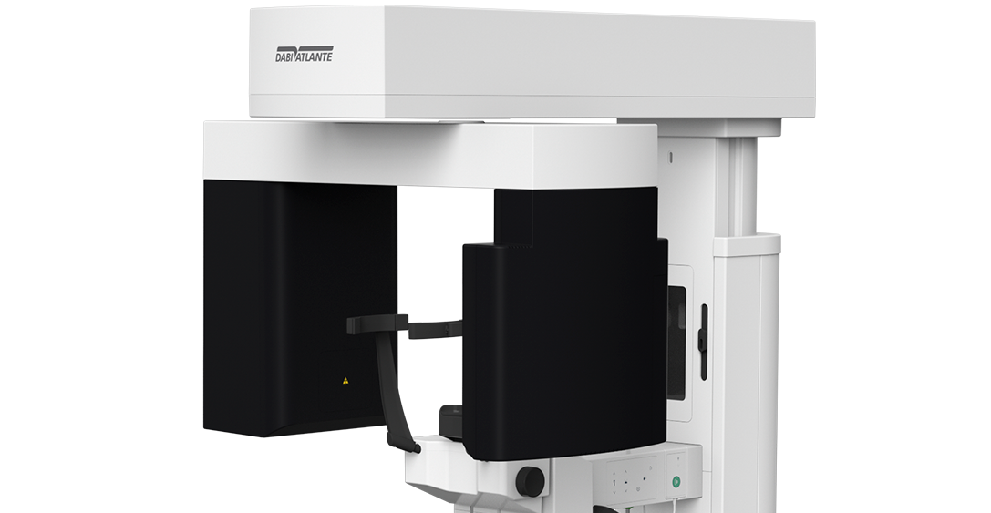
Stability and usability
Eagle Edge has new head positioners with 4 support points for better patient stability during exams. Simple to use and easy to position, the set of head positioners was designed to facilitate the clinical routine, making the execution in sequence faster.
DICOM Server
The Dicom Send tool of the Eagle software allows the instantaneous sending of the images generated by the equipment to image storage and sharing systems in physically different locations.
Reduction of metallic artifacts
Eagle Edge features options with selectable processing levels to correct deformations of gutta-percha, implants and/or wide prostheses and metal restorations, in addition to automatic metal reduction. This feature also allows image reprocessing for a better diagnosis without the need to generate new exposure to the patient.